Monday, July 18, 2016

ANGIOGRAPHY--A WHOLEHEARTED AID FOR HUMAN BEING

Angiography or arteriography is a medical imaging technique used to visualize the inside, or lumen, of blood vessels and organs of the body, with particular interest in the arteries, veins, and the heart chambers. This is traditionally done by injecting a radio-opaque contrast agent into the blood vessel and imaging using X-ray based techniques such asfluoroscopy.
The word itself comes from the Greek words ἀνγεῖον angeion, "vessel", and γράφειν graphein, "to write" or "record". The film or image of the blood vessels is called an angiograph, or more commonly, an angiogram. Though the word itself can describe both an arteriogram and a venogram, in its everyday usage, the terms angiogram and arteriogram are often used synonymously, whereas the term venogram is used more precisely.
The term angiography is strictly defined as based on projectional radiography; however, the term has been applied to newer vascular imaging techniques such as CT angiography and MR angiography. The term isotope angiography has also been used, although this more correctly is referred to as isotope perfusion scanning.
History
The technique was first developed in 1927 by the Portuguese physician and neurologist Egas Moniz at the University of Lisbon to provide contrasted x-ray cerebral angiography in order to diagnose several kinds of nervous diseases, such as tumors, artery disease and arteriovenous malformations. He is usually recognized as one of the pioneers in this field. Moniz performed the first cerebral angiogram in Lisbon in 1927, and Reynaldo Cid dos Santos performed the first aortogram in the same city in 1929. With the introduction of the Seldinger technique in 1953, the procedure became markedly safer as no sharp introductory devices needed to remain inside the vascular lumen.

History

The technique was first developed in 1927 by the Portuguese physician and neurologist Egas Moniz at the University of Lisbon to provide contrasted x-ray cerebral angiography in order to diagnose several kinds of nervous diseases, such as tumors, artery disease and arteriovenous malformations. He is usually recognized as one of the pioneers in this field. Moniz performed the first cerebral angiogram in Lisbon in 1927, and Reynaldo Cid dos Santos performed the first aortogram in the same city in 1929. With the introduction of the Seldinger technique in 1953, the procedure became markedly safer as no sharp introductory devices needed to remain inside the vascular lumen.

Technique

Depending on the type of angiogram, access to the blood vessels is gained most commonly through the femoral artery, to look at the left side of the heart and at thearterial system; or the jugular or femoral vein, to look at the right side of the heart and at the venous system. Using a system of guide wires and catheters, a type ofcontrast agent (which shows up by absorbing the x-rays), is added to the blood to make it visible on the x-ray images.
The X-ray images taken may either be still images, displayed on an image intensifier or film, or motion images. For all structures except the heart, the images are usually taken using a technique called digital subtraction angiography or DSA. Images in this case are usually taken at 2 – 3 frames per second, which allows theinterventional radiologist to evaluate the flow of the blood through a vessel or vessels. This technique "subtracts" the bones and other organs so only the vessels filled with contrast agent can be seen. The heart images are taken at 15–30 frames per second, not using a subtraction technique. Because DSA requires the patient to remain motionless, it cannot be used on the heart. Both these techniques enable the interventional radiologist or cardiologist to see stenosis (blockages or narrowings) inside the vessel which may be inhibiting the flow of blood and causing pain.
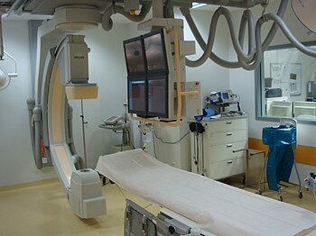
Catheterization Lab
Catheterization in selective angiography

Uses

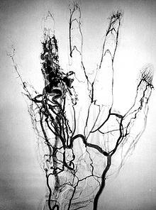
Finger angioma seen on angiogram

Coronary angiography

Main article: Coronary angiography
One of the most common angiograms performed is to visualize the blood in the coronary arteries. A long, thin, flexible tube called a catheter is used to administer the X-ray contrast agent at the desired area to be visualized. The catheter is threaded into an artery in the forearm, and the tip is advanced through the arterial system into the major coronary artery. X-rayimages of the transient radiocontrast distribution within the blood flowing inside the coronary arteries allows visualization of the size of the artery openings. Presence or absence of atherosclerosis or atheroma within the walls of the arteries cannot be clearly determined. See coronary catheterization for more detail.
To detect coronary artery disease, Computed Tomography (CT) Scan is better than Magnetic Resonance Imaging (MRI). The sensitivity and specificity between CT and MRI were (97.2 percent and 87.4 percent) and (87.1 percent and 70.3 percent), respectively. Therefore, CT (mainly multislice CT) is more accepted, more widely available, more favored by patients, and more economic. Moreover, CT requires shorter breath-hold time than MRI.

Microangiography

Microangiography is commonly used to visualize tiny blood vessels.

Neuro-vascular angiography

Another increasingly common angiographic procedure is neuro-vascular digital subtraction angiography in order to visualise the arterial and venous supply to the brain. Intervention work such as coil-embolisation of aneurysms and AVM gluing can also be performed.

Peripheral angiography

Angiography is also commonly performed to identify vessel narrowing in patients with leg claudication or cramps, caused by reduced blood flow down the legs and to the feet; in patients with renal stenosis (which commonly causes high blood pressure) and can be used in the head to find and repair stroke. These are all done routinely through the femoral artery, but can also be performed through the brachial or axillary (arm) artery. Any stenoses found may be treated by the use ofatherectomy.

Post mortem CT angiography for medicolegal cases

Post mortem CT angiography for medicolegal cases is a method initially developed by the Virtopsy group. Originating from that project, both watery and oily solutions have been evaluated.
While oily solutions require special deposition equipment to collect waste water, watery solutions seem to be regarded as less problematic. Watery solutions also were documented to enhance post mortem CT tissue differentiation whereas oily solutions were not. Conversely, oily solutions seem to only minimally disturb ensuing toxicological analysis, while watery solutions may significantly impede toxicological analysis, thus requiring blood sample preservation before post mortem CT angiography

Complications

After an angiogram, a sudden shock can cause a little pain at the surgery area, but heart attacks and heart strokes usually don't occur, as they may in bypass surgery.
Cerebral angiography

Major complications in cerebral angiography such as in Digital subtraction angiography or contrast MRI are also rare but include stroke, an allergic reaction to theanaesthetic other medication or the contrast medium, blockage or damage to one of the access veins in the leg, or thrombosis and embolism formation. Bleeding orbruising at the site where the contrast is injected are minor complications, delayed bleeding can also occur but is rare.

Additional risks

The contrast medium that is used usually produces a sensation of warmth lasting only a few seconds, but may be felt in a greater degree in the area of injection. If the patient is allergic to the contrast medium, much more serious side effects are inevitable; however, with new contrast agents the risk of a severe reaction are less than one in 80,000 examinations. Additionally, damage to blood vessels can occur at the site of puncture/injection, and anywhere along the vessel during passage of the catheter. If digital subtraction angiography is used instead, the risks are considerably reduced because the catheter does not need to be passed as far into the blood vessels; thus lessening the chances of damage or blockage.

Sunday, July 17, 2016

RADIOGRAPHY TECHNOLOGIST --A JOB WITH CONCERNS?

Radiographers, also known as Radiologic TechnologistsDiagnostic RadiographersMedical Radiation Technologists are Healthcare Professionals who specialise in the imaging of human anatomy for the diagnosis and treatment of pathology. Radiographers are infrequently, and almost always erroneously, known as X-Ray Technicians. In countries which use the title radiologic technologist they are often informally referred to astechs in the clinical environment; this phrase has emerged in popular culture such as television programmes.
Radiographers work in both public and private healthcare and can be physically located in any setting where appropriate diagnostic equipment is located, most frequently in hospitals. Their practice varies country to country and can even vary between hospitals in the same country.
Radiographers are represented by a variety of organisation worldwide, the International Society of Radiographers and Radiologic Technologists  aims to give direction to the profession as a whole through collaboration with national representative bodies.

Risks

  • Epidemiological studies indicate that Radiographers employed before 1950 are at increased risk of leukemia and skin cancer, most likely due to the lack of use of radiation monitoring and shielding.
  • Ionising radiation, used in a variety of imaging procedures, can damage cells. Lead shields are used on the patient and by the Radiographer to reduce exposure by shielding areas that do not need to be imaged from the radiation source. While lead is highly toxic, the shields used in medical imaging are coated to prevent lead exposure and are regularly tested for integrity.
  • Radiographers who develop x-ray films are exposed to the various chemical hazards such as sulfur dioxide, glutaraldehyde, and acetic acid. These agents can cause asthma and other health issues.
  • Theoretically, the strong static magnetic fields of MRI scanners can cause physiological changes. After a human neural cell culture was exposed to a static magnetic field for 15 minutes, changes in cell morphology occurred along with some modifications in the physiological functions of those cells. However, these effects have not yet been independently replicated or confirmed, and this particular study was performed in vitro.
  • Ultrasound imaging can deform cells in the imaging field, if those cells are in a fluid. However, this effect is not sufficient to damage the cells.
  • As with any allied health professional, exposure to infectious diseases is likely, and use of Personal Protective Equipment (PPE) and infection control precautions must be employed to reduce the risk of infection.

Saturday, July 16, 2016

THE AMAZING EVOLUTION OF RADIOLOGICAL TECHNOLOGY

The history of radiology began in 1895 when a German physicist, named Wilhelm Conrad Roentgen, discovered what he called a “new kind of ray.”
He found that these rays could pass through human tissue but not bones and metal. One of his first experiments was taking an X- ray of his wife’s hand, as seen below.
Since these early X-rays, there have been many milestones in the field of radiology, including:
1942: First recorded use of ultrasound for medical diagnosis
1971: First CT scan is taken at Atkinson Morley’s Hospital, in London, England
1977: First full body, human MRI image taken
1970’s: Use of real time ultrasound makes it the common practice for medical diagnosis
While medical imaging technology has greatly advanced in 118 years, the ways in which we interact with those images has remained largely unchanged.
 Most patients still carry around bulky files of their images from doctor to doctor with the risk of getting lost or damaged.
With Purview, patients can easily store and share their medical images online. Patients and doctors all over the country can then access these digital images at anytime.
Direct access and control to digital medical images is the next evolution in radiology. We’re revolutionizing the industry in ways Wilhelm Roentgen couldn’t possibly imagine.
Certainly, we have come a long way in 121 years. We have CT scanners, MRI scanners. Those imagining modalities capture hundreds of medical images during a given procedure. Doctors are able to look at those images in 2D and in 3D. They are able to look at them from anywhere.
What hasn’t changed is how patients engage with those medical images. Patients still carry a sheet of film from doctor to doctor. Typically, doctors look at those sheets of film on a light box and the patient is told to go home and store them between their box spring and their mattress.
Purview is trying to take medical imaging to the next level. We are trying to democratize health care for patients. We are trying to make health records portable and accessible. So that X-ray that you only had one copy of 121 years ago, or even last week, now you have a digital copy in the cloud that you can easily share with health care providers anywhere, anytime.

I.V.P. still useful for us

An intravenous pyelogram (IVP) is an x-ray examination of the kidneys, ureters and urinary bladder that uses iodinated contrast material injected into veins.
An x-ray (radiograph) is a noninvasive medical test that helps physicians diagnose and treat medical conditions. Imaging with x-rays involves exposing a part of the body to a small dose of ionizing radiation to produce pictures of the inside of the body. X-rays are the oldest and most frequently used form of medical imaging.
When contrast material is injected into a vein in the patient's arm, it travels through the blood stream and collects in the kidneys and urinary tract, turning these areas bright white on the x-ray images. An IVP allows the radiologist to view and assess the anatomy and function of the kidneys, ureters and the bladder.

What are some common uses of the procedure?

An intravenous pyelogram examination helps the radiologist assess abnormalities in the urinary system, as well as how quickly and efficiently the patient's system is able to handle fluid waste.
The exam is used to help diagnose symptoms such as blood in the urine or pain in the side or lower back.
The IVP exam can enable the radiologist to detect problems within the urinary tract resulting from:
  • kidney stones
  • enlarged prostate
  • tumors in the kidney, ureters or urinary bladder
  • scarring from urinary tract infection
  • surgery on the urinary tract
  • congenital anomalies of the urinary tract

How should I prepare?

Your doctor will give you detailed instructions on how to prepare for your IVP study.
You will likely be instructed not to eat or drink after midnight on the night before your exam. You may also be asked to take a mild laxatives (in either pill or liquid form) the evening before the procedure.
You should inform your physician of any medications being taken and if there are any allergies, especially to iodinated contrast material . Also inform your doctor about recent illnesses or other medical conditions.
You may be asked to remove some or all of your clothes and to wear a gown during the exam. You may also be asked to remove jewelry, removable dental appliances, eye glasses and any metal objects or clothing that might interfere with the x-ray images.
Women should always inform their physician and x-ray technologist if there is any possibility that they are pregnant. Many imaging tests are not performed during pregnancy so as not to expose the fetus to radiation. If an x-ray is necessary, precautions will be taken to minimize radiation exposure to the baby. 

What does the equipment look like?

The equipment typically used for this examination consists of a radiographic table, one or two x-ray tubes and a television-like monitor that is located in the examining room. Fluoroscopy, which converts x-rays into video images, is used to watch and guide progress of the procedure. The video is produced by the x-ray machine and a detector that is suspended over a table on which the patient lies.

How does the procedure work?

X-rays are a form of radiation like light or radio waves. X-rays pass through most objects, including the body. Once it is carefully aimed at the part of the body being examined, an x-ray machine produces a small burst of radiation that passes through the body, recording an image on photographic film or a special detector.
In an IVP exam, an iodine-containing contrast material is injected through a vein in the arm. The contrast material then collects in the kidneys, ureters and bladder, sharply defining their appearance in bright white on the x-ray images.
X-ray images are typically stored as digital images in an electronic archive. However, if needed, a hard film copy (similar to a photograph) or a CD-ROM disk can be made. These stored images are easily accessible and may be compared to current or prior x-ray images for diagnosis and disease management.

How is the procedure performed?

This examination is usually done on an outpatient basis.
You will lie on the table and still x-ray images are taken. The contrast material is then injected, usually in a vein in your arm, followed by additional still images. The number of images taken depends on the reason for the examination and your anatomy.
You must hold very still and may be asked to keep from breathing for a few seconds while the x-ray picture is taken to reduce the possibility of a blurred image. The technologistwill walk behind a wall or into the next room to activate the x-ray machine.
As the contrast material is processed by the kidneys, a series of images is taken to determine the actual size of the kidneys and to image the urinary tract in action as it begins to empty. The technologist may apply a compression band around the body to better visualize the urinary structures.
When the examination is complete, you may be asked to wait until the radiologist determines that all the necessary images have been obtained.
An IVP study is usually completed within an hour. However, because some kidneys function at a slower rate, the exam may last up to four hours.

Saturday, February 7, 2009

BRAIN METASTASES(CANCER) & RADIOGRAPHY


In case of various type of brain metastases there is no other option than radiography, specially MRI & CT scan. These can be easily diagnosed various type of tumors in brain & then one can look for any other primary lesion in body. It is very essential to take a right decision to go for CT scan of brain or for MRI because in some cases MRI is more effective than CT scan & in some cases CT scan is more acceptable.

Using a contrast injection that is non ionic radiopaque (76%Iodine) contrast media we can easily diagnosed the type or name of tumors in brain.


MRI-basic principles

Clinical Magnetic Resonance Imaging (MRI) uses the magnetic properties
of hydrogen and its interaction with both a large external magnetic field
and radio waves to produce highly detailed images of the human body. In
this first module, we will discuss some basic principles of magnetism, the
magnetic properties of the hydrogen nucleus, and its interaction with the
externally applied magnetic field (B0).
In its early days, MRI was known as NMR. This stands for Nuclear Magnetic
Resonance. Although the name has changed (primarily due to the
negative connotation of the word “nuclear”), the basic principles are the
same. We derive our images from the magnetic resonance properties of
nuclear particles (specifically hydrogen).
In order to perform MRI, we first need a strong magnetic field. The field
strength of the magnets used for MR is measured in units of Tesla. One
(1) Tesla is equal to 10,000 Gauss. The magnetic field of the earth is
approximately 0.5 Gauss. Given that relationship, a 1.0 T magnet has a
magnetic field approximately 20,000 times stronger than that of the
earth. The type of magnets used for MR imaging usually belongs to one of
three types; permanent, resistive, and superconductive.
A permanent magnet is sometimes referred to as a vertical field magnet.
These magnets are constructed of two magnets (one at each pole).

The patient lies on a scanning table between these two plates.
Advantages of these systems are: 1) Relatively low cost, 2) No electricity
or cryogenic liquids are needed to maintain the magnetic field, 3) Their
more open design may help alleviate some patient anxiety, 4) Nearly nonexistant
fringe field. It should be noted that not all vertical field magnets
are permanent magnets.
Resistive magnets are constructed from a coil of wire. The more turns to
the coil, and the more current in the coil, the higher the magnetic field.
These types of magnets are most often designed to produce a horizontal
field due to their solenoid design (figure 2).
As previously mentioned, some vertical field systems are based on
resistive magnets. The main advantages of these types of magnets are:
1) No liquid cryogen, 2) The ability to “turn off” the magnetic field, 3)
Relatively small fringe field.
resistive system:

DIAGNOSTIC RADIOGRAPHY-playing main role in medical sector

Diagnostic radiography is a very common word for us now. More or less all of us have experienced it any of time in our life. X-RAY, CT SCAN, MRI are now not very unknown words for us. Yes, these all are nothing but radiography or radiology. Now these are obvious part of our life, saving our life. If X-RAY is the mother of radiography, then CT scan & MRI come for next generation. Though CT scan based on x-ray, but MRI is not based on it. CT scan represents Computed Tomography while MRI stands for Magnetic Resonance Imaging & no relation with x-ray. Every aspect of these three aids have their own unique applications. While x-ray is very effective for some cases, then CT scan has also some specialties & has great advantages in diagnosis for many disease. Then MRI comes & proves itself as a great fusion of technology & medical science. Radiology is an art & the picture has to be technically & photographically good to be diagnostic.